
Anatomically, the heart is located in the anterior thoracic cavity so when a person faces forward toward an X-ray machine, the heart is closer to the X-ray machine and farther from the film behind it. Contrarily, what happens if you hold the pen as far as you can away from the flashlight, so that it sits directly in front of the wall? In this case, the shadow is much smaller, and is a much more accurate representation of the pen’s true size.ĭo you see now the relevance of this example? The same thing that happens to the pen’s shadow when you hold it closer to the flashlight and farther from the wall happens to the picture of the heart created by the rays in an AP view. If you hold the pen directly in front of the flashlight, is the shadow big or small? In this case, the shadow formed by the pen is large, and much larger than the pen’s true size. Now imagine holding a pen in front of that light, so that the pen casts a shadow onto the wall. Imagine you are holding a flashlight, pointing it so that the circle of light appears against a white wall a foot away from you. Let’s take a second to try to understand why it is that the heart appears bigger than normal on AP studies. To take left lateral films, the patient stands or sits upright with his or her arms raised and turns 90 degrees so that the left side faces the receptor this allows the X-ray beams to travel from the emitter through the patient from right to left to the receptor on the other side. the lungs are filled with as much air as the patient is capable of inhaling). PA films are the standard: the patient stands or sits upright approximately 6 feet in front of the beam source and faces the receptor on the other side, with the X-ray taken while the patient is maximally inspiring (i.e. These terms refer to the patient’s position and therefore tell you the direction that the X-ray beam travels through the body to the receptor. When presented with a chest X-ray, the first thing one should do is try to determine the view, that is, the positions of the patient and machine and thus the trajectory of the rays relative to the patient. Penetration: the vertebrae behind the heart are barely visible, and the diaphragm can be traced up until reaching the edge of the spine.Įdges, effusions, extrathoracic soft tissues Inspiration: counting the posterior ribs visible in the lung fields Rotation: compare the positions of the left and right medial clavicular joints to the spinous processes in the more central aspect of the image Lateral: patient stands or sits upright with his or her arms raised and turns 90 degrees so that the left side faces the receptorĪnteroposterior: patient lies down on top of the receptor, such that the X-ray beam travels through the patient from front to back Posteroanterior (standard): patient stands or sits upright approximately 6 feet in front of the beam source and faces the receptor.
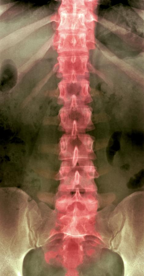
#Normal spine xray how to#
Key facts about how to read chest X-rays Determining the view In this image, air is black, bones are white, and the rest of the tissues fall on a spectrum in between.

This differential absorption allows for the creation of an image on radiographic film. Some of these rays are absorbed more than others depending on the tissues through which they travel. X-rays are emitted by a machine, travel through the patient, and are picked up by a receptor on the other side of the patient.

It is almost always the first imaging study ordered to evaluate for pathologies of the thorax, although further diagnostic imaging, laboratory tests, and additional physical examinations may be necessary to help confirm the diagnosis. X-ray of the chest (also known as a chest radiograph) is a commonly used imaging study, and is the most frequently performed imaging study in the United States.


 0 kommentar(er)
